-
About
-
Mission
-
Faculty
-
Projects
Daphnia Swarming
EEG/fMRI Correlation
Eye-Target Synchrony
Synchronization in Crayfish
Behavioral Stochastic Resonance
Visual Stochastic Resonance
-
Students
Kaushalya Premachandra
Dawn King
Adam Scott
-
Alumni
Gabor Balazsi
Alexander Neiman
Jorge Brea
Nathan Dees
Roxana Contreras
Douglas Brumm
Ricardo Garcia
David F. Russell
Kevin Dolan
Jennifer Simonotto
Enrico Simonotto
Xing Pei
Winfried Wojtenek
Anke Ordemann
Oliver Weihberger
Sallie Breite
Nicola Jung
Daisuke Takeshita
-
Post Docs
Corey Maley, Post Doctoral Fellow
-
Certificate in Neuroscience
-
Contact Us

EEG/fMRI Correlation
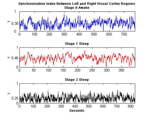
Using simultaneously recorded Electroencephalographic (EEG) and functional Magnetic Resonance Image (fMRI) data of subjects resting quietly with their eyes closed (awake) and secondly sleeping (not deeply), this team is looking fervently for a connection between the two datasets to more fully utilize their respective qualities. EEG data has temporal resolution in the millisecond range, but very poor spatial resolution, while fMRI has great 3D spatial resolution, however sadly slow temporal resolution. A more complete picture of the working brain would incorporate the advantages of both modalities.
The team has employed numerous linear and nonlinear measures to better understand the data, most notably cross-correlation measures, stochastic phase synchronization measures, coherence measures, and evolution map approach measures. Information theory and power measurements have also been utilized to try to make the connection, and perhaps distinguish between awake subjects, and drowsy/sleepy subjects. The focus has been on the left and right visual cortex and the left and right dorsolateral prefrontal cortex, and also the frequency ranges delta, theta, and alpha.
Moving forward, the group will begin to analyze electrocorticographic (ECoG) data of more deeply sleeping subjects and also will be working on capturing visually evoked potential (VEP) data in EEG and fMRI. New analysis methods will be incorporated also including wavelet analysis and parallel factor (PARAFAC) analysis.
Collaboration with Linda Larson-Prior of the Electrical and Neuro-Optical Imaging Lab at Washing University School of Medicine. Research updates coming soon.
