 Dimpling
Navigation bar: [intro] [task] [process] [vEM] [resources] [evaluation] [conclusion] [credits] "Go back" to return.
The prefix "nano" in the word nanoworld means a billionth (10-9). When exploring the nanoworld, one deals with various structures of matter having dimensions of the order of a billionth of a meter. Although windows into the nanoworld have begun to open only a few decades ago, structures of nanometer dimensions have always existed on Earth. One of the major instruments used to examine matter at nanoscale dimensions is the electron microscope, which uses electrons instead of visible light to image samples. An extensive electron microscopy facility is located at UM-St. Louis in the Center for Molecular Electronics' Scanned Tip and Electron Image Lab. When you begin this exercise, you will see your specimen in the specimen mounting area of an actual atomic-resolution transmission electron microscope (TEM). You will explore this specimen, and look for and make measurements and observations on some nanoworld objects of interest to chemists, physicists, and biologists.
As a nano-detective for this exercise, your prime instrument will be a simulated or 'virtual' electron microscope (vEM). The objects of interest are located on the surface of a 3-mm silicon-specimen disc. Look around and you will find them! These empirical observation challenges will bridge the gap between your classroom and 'real world' experiences, and enhance the development of your critical observation and communication skills. Have you ever wondered how small atoms are and how much space there is between two adjacent atoms?
Use the vEM to zoom down for a closer look at what you can find on the 3-mm specimen. Please read the notes under 'process' and 'evaluation' before starting to work. You may save your work and return to it later. It is recommended that you place your work log, which is like your 'virtual lab notebook' in the 'comments' part of the Assignment. The rest of your work (introduction, method, data, interpretation, and uncertainties) should be entered into a Microsoft Word document and attached when you are ready to submit your assignment. Submission of your assignment will email it directly to me, but only do this when you are convinced that you are done. Handwritten reports will not be accepted.
There is not one rigid set of correct answers to this set of tasks, different students will find different things and end up with different answers within a reasonable range.
As a nano-detective, you will need to develop your own answers to the following questions:
The picture below should give you some ideas about the relative size of the specimen that you will be exploring. This is a typical "self-supporting" transmission electron microscope specimen. Specimens requiring support (e.g. nanoparticles) typically use a 3[mm] TEM grid instead.
The 3-mm diameter disc is located at the tip of the tweezer if you haven't noticed it. Now let's further examine the specimen by using the magnifying glass (use your mouse to grab its handle with a hand icon). The magnifying glass didn't help much, did it? For those with urgent curiosity, click on the specimen when it is visible under the magnifying glass. This will transport the specimen into a separately-windowed "virtual electron microscope" for further examinations, but more on that later. Now let's learn how the tiny specimen is made.
Specimen Preparation
Disk cutting and thinning
The specimen has to fit into the tight space of a magnetic lens polepiece, and be thin enough in regions of interest to let electrons to pass through (i.e. well under 1 micron thick). Normally an ultrasonic disk-cutter is used to cut a large sample into a flat disk of 3 millimeters in diameter. This disk is ground down with diamond powder to a uniform thickness of about 0.1 millimeters.
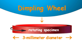
Dimpling
In this process, the disk is ground futher with a so-called dimpler which makes a slight hollow (dimple) in the center of the specimen. In this picture only the lower part of the dimpling wheel used for grinding is drawn. The disk is rotating all the time to make the dimple in the center uniform.
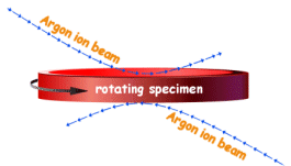 Ion-milling
The dimpled disk is put into a machine that is called an ion-mill. Two beams of argon ions (shown here in blue) are directed at the disk which is once again rotating. These beams slowly erode material from the top and bottom of the disk. The ion-milling is continued until a hole appears approximately in the center of the disk.
Begin Your Explorations
In order to begin your detective work, you need to learn how to use the virtual electron microscope. Don't be scared, the vEM is easy to operate. Just follow these simple rules:
Now you are fully equipped. Let the fun begin!
* Computers often let you capture screen images to a clipboard (here are some instructions if you are running Windows) for pasting into a graphics or word processing program.
Reporting Results: Show Your Discovery
You are to write a scientific diary and report about your adventures as a nano-detective. We recommend that your report consist of 6 sections:
1). Work Log: This is a simple diary of project activity with entries of the form "date-time/what happened".
2). Introduction: This section describes what you can find out about the challenge before actually doing the experiment. Here references to external sources are crucial.
3). Method: Describe here how you took data, with enough detail that another student in the course could repeat your experiment and get approximately the same results.
4). Data/Observations: This section reports the actual measurements that you made. These might be typed directly into the text, or simply summarized in the text and provided separately in spreadsheet or image attachments.
5). Interpretation/Theory: In this section, you make the connection between your observations and the objective of the nanoworld challenge. What conclusions are possible, based on your observations?
6). Assumptions/Uncertainties: Here is where you address the strength of your
interpretation. What assumptions are involved, and how likely are they to be
valid? If you have quantitative conclusions, provide evidence concerning the
expected numerical error in those conclusions. This might also be a good place
to mention possible future work.

Get to know microscopic objects that you may bump into on the virtual specimen:
Additional resources:
Your final grade for this WebQuest activity will be assessed using a 40-point rubic* below that details what you will need to accomplish to earn points. Be sure to refer back to it often to help you know how you are doing as you progress in the project.
|
||||||||||||||||||||||||||||||||||||||||||||||||||
Understanding the microscopic world of atoms requires having a good sense of scale to compare the visual world to that of the tiny atom. Nature has proven to be a most skilled architect on this smallest size-scale of both biology's gene-driven, and technology's idea-driven, innovation. The size of atoms is one characteristic that is quite important for understanding their behavior. I hope that this activity has also let you hone your skills as a nano-detective, and provided a sense of the many levels of natural phenomena that separate us from the atoms that make us up.
This page is
http://www.umsl.edu/~fraundor/nanowrld/live3Dmodels/chemapp.htm.
Suggestions toward improvement, and correction, are invited.
The page was put together with material and guidance from
Keith Stine in Chemistry and BioChemistry at UM-StL, for
an application in his classroom. The text page numbers
refer to the course text by J. E. McMurry and R. C. Fay,
Chemistry, 4th Edition, Prentice-Hall, Inc. (2004).
The person responsible for
errors is P. Fraundorf. Two acknowledgements are particularly
important. Former grad student Noom Pongkrapan adapted the ideas used
here into webQuest format, added less intimidating prose, put together
the specimen prep figures, and developed the Macromedia Flash "magnifying
glass" illustration. Martin Kraus provided the Live3D
applet, and helped adapt it to this application with
significant modifications. This site is hosted by the Department of
Physics and Astronomy (and Center for Molecular Electronics)
at UM-StLouis.
Mindquilts site
page requests est. around 2000/day
hence more than 500,000/year. Page requests to a stat-counter
linked subset of pages since 4/7/2005:
.